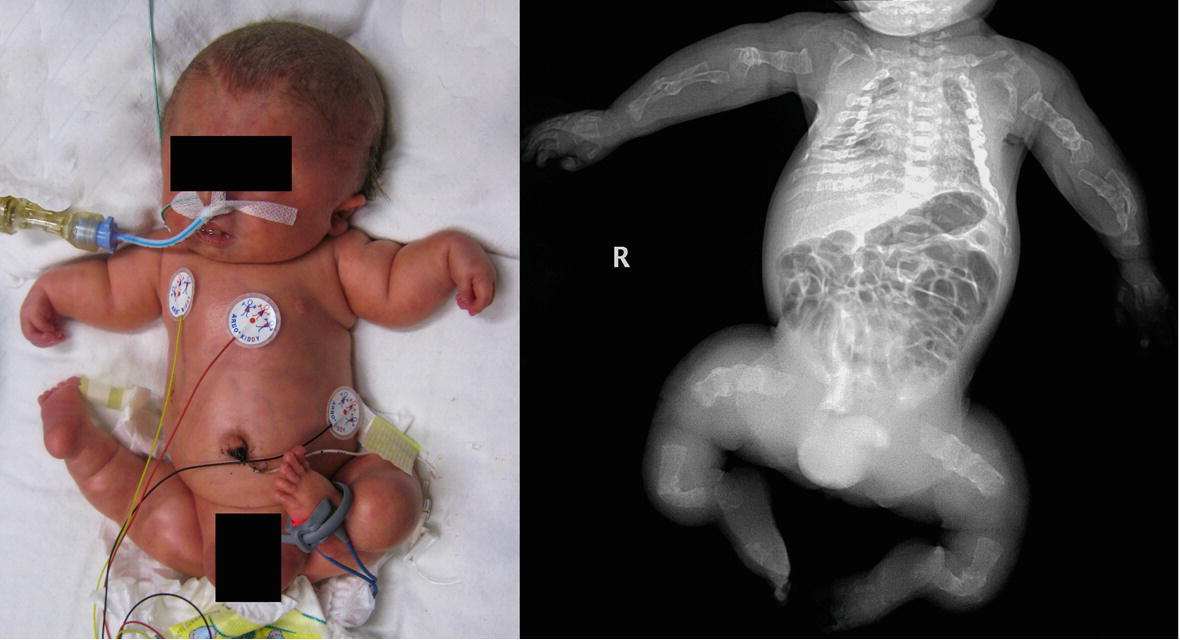

Taitz estimated the incidence of OI type IV without features of the disease or a family history in infants presenting under 1 year of age to be between 1 in 1 million and 1 in 3 million births ( 20), but it is unclear whether this figure is still applicable in the era of genetic testing.įigure 9.4 OI type IV presenting with suspected abuse.

OI type IV is rare, constituting less than 5% of all cases of OI.

At times, distinction from type I is difficult, especially in early childhood, and some authors feel that these type IV cases actually belong to the first type. The fracture onset can be prenatal, and again, demineralization is generally appreciable on high-quality radiographs ( Figs. Radiographically, there may be evidence of platybasia with an “occipital overhang.” Short stature is commonly present (40%) even in the newborn period. This form of OI is also associated with bluish or grayish sclerae. The craniofacial relationships are often abnormal, with a triangular face, biparietal widening, and a broad forehead. OI type IV has a phenotype similar to type I except for several distinctive features. C, AP radiograph of the pelvis and femurs at nine months of age shows generalized demineralization. A, B, AP and lateral radiographs at seven months of age show a metadiaphyseal insufficiency fracture (arrows) of the left tibia with generalized demineralization and cortical thinning. No genetic defect discovered at time of diagnosis. Figure 9.2 Clinically confirmed OI type I, type I collagen analysis negative.


 0 kommentar(er)
